NEUROPHET tES LAB can makes fully automated brain segmentation
Description

NEUROPHET tES LAB is the powerful research software for the computational study to predict and analyze the simulation effect of tDCS/tACS.
It can analysis the effect field (EF) distribution and magnitude through the finite element method (FEM) in a personalized brain model. It can be applied to find the optimal stimulation parameters. All steps are automated to reduce the inconvenience of manual operations and the operation time from hours to minutes. It can be applied in various fields such as neurology, medical imaging, neuromodulation, rehabilitation medicine, psychiatry, and sports science.
– Fully automatic segmentation
Using T1-weighted MRI data, the AI engine segments it automatically. Even skin and skulls that are crucial factors for tDCS can be segmented.
– Personalized 3D model generation
The automatic brain modeling engine generates a 3D individual brain model based on the MRI data segmentation. This will increase accuracy in positioning electrodes and result in better stimulation effects.
– Electrode customizing
Users can customize electrodes in sizes, shapes, numbers and position with just a few clicks. Selecting types of stimulation by tDCS/tACS/tDCS Optimization/10-20 System-based Optimization is also available.
– 10-20 system support
After specify four landmarks, User can set experimental conditions using the 10-20 system on various channels. (16ch/32ch/64ch)
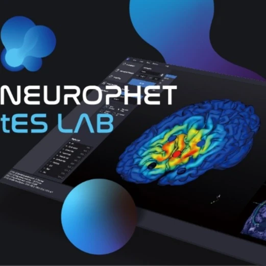
– Stimulation effect prediction and result analysis
The high-speed simulation engine visualizes the generated stimulation effect in the brain quickly and accurately. It allows users to confirm the intensity and activation/inhibition effects on the stimulation parameters.
– Stimulation optimization
The powerful simulation engine determines conditions that effectively stimulate the brain’s stimulation target.
Supported MRI data format
Image format | NIfTI – 1 (file extension : .nii) |
Type | Structural MRI (T1-weighted) |
Slice thickness (Spacing) | Coronal ≤ 1.0 mm Sagittal ≤ 1.0 mm Axial ≤ 1.6 mm |
Field Strength (Tesla) | 1.5T 3.0T |
Validated devices and protocols of MRI
Company | Model | Protocol |
GE | Signa HDxt 1.5T | |
Philips | Intera 1.5T | MPRAGE |
Intera 3T Ingenia 3T | TFE | |
Siemens | Skyra 3T Verio 3T | MPRAGE |
– Hardware Specification
NEUROPHET leads the advanced neuroscience technologies and contributes to conquering diverse brain diseases for a better quality of life. We develop practical AI medical solutions enabling to apply them to medical and research fields.
The advanced brain segmentation and modeling technology ‘AI-powered NEUROPHET SegEngine’ was begun from ‘The next generation neuronavigation system’ project of GIST, one of the major research institutes in Korea, in 2010. Our unparalleled technology is applied to the various NEUROPHET’s products such as the neuromodulation research software and the neurological diagnosis software.
As a global healthcare enterprise, NEUROPEHT is dedicated to expanding our business throughout the world and maintaining a reliable reputation by improving technologies.
Additional information
| Brand Name: | NEUROPHET |
|---|---|
| Model Number: | tES LAB |
| Place of Origin: | South Korea |
| Instrument classification: | Class II |
| CODE: | SVO19 |